Banner

Histology Laboratory


Images


نوع بافت

عكسها

اپيتليوم و انواع آن

عكس

بافت

همبند

استخوان

عكس

غضروف

             عكس

تاندون

عكس

بافت همبند سست

عكس

چربي

عكس

خون وسلولها

عكس

دستگاه گردش خون

عكس

دستگاه عضلاني

عكس

دستگاه ايمني

-

دستگاه گوارش و غدد ضميمه

عكس    عكس   

دستگاه تنفس

عكس

پوست

عكس

دستگاه ادراري

عكس

دستگاه غدد درون ريز

عكس

دستگاه عصبي و حسي

عكس    عكس 

دستگاه توليد مثل مرد

عكس

دستگاه توليد مثل زن

عكس


EPITHELIAL TISSUE.. 18

CONNECTIVE TISSUE.. 22

BONE.. 22

CARTILAGE, DENSE FIBROUS CONNECTIVE TISSUE (TENDON) 25

LOOSE FIBROUS CONNECTIVE TISSUE.. 27

ADIPOSE.. 27

BLOOD.. 28

CIRCULATORY SYSTEM... 30

MUSCLE TISSUE.. 35

NERVOUS TISSUE.. 37

SENSE ORGANS. 50

DIGESTIVE SYSTEM... 56

ESOPHAGUS, STOMACH.. 56

INTESTINE.. 59

LIVER, PANCREAS. 65

TONGUE, TOOTH, TOOTH DEVELOPMENT. 69

ENDOCRINE SYSTEM... 72

PITUITARY GLAND (HYPOPHYSIS), PINEAL GLAND (EPIPHYSIS) 72

THYROID GLAND, PARATHYROID LAND, ADRENAL GLAND.. 74

RESPIRATORY SYSTEM... 77

SKIN.. 83

URINARY SYSTEM... 87

REPRODUCTIVE SYSTEM... 90

MALE REPRODUCTIVE SYSTEM  TESTIS. 91

FEMALE REPRODUCTIVE SYSTEM... 99


EPITHELIAL TISSUE


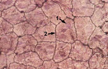
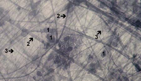
MESOTHELIUM (SIMPLE SQUAMOUS EPITHELIUM)
view from surface
     Stained with silver nitrate

1 - nucleus of cell
2 - cell borders


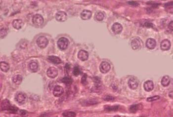
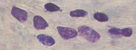
SIMPLE SQUAMOUS EPITHELIUM
     Stained with haematoxylin and eosin
Nuclei of the epithelial cells are shown with an arrow


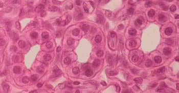
SIMPLE CUBOIDAL EPITHELIUM
     Stained with haematoxylin and eosin


SIMPLE CUBOIDAL EPITHELIUM
     Stained with haematoxylin and eosin


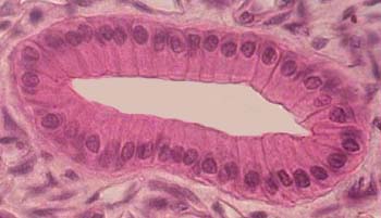
SIMPLE COLUMNAL EPITHELIUM
     Stained with haematoxylin and eosin


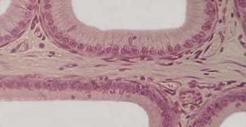
SIMPLE PSEUDOSTRATIFIED COLUMNAR EPITHELIUM
     Stained with haematoxylin and eosin


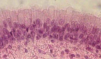
SIMPLE PSEUDOSTRATIFIED COLUMNAR CILIATED EPITHELIUM
     Stained with haematoxylin and eosin


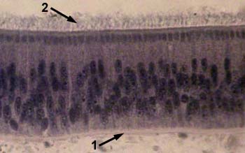
SIMPLE PSEUDOSTRATIFIED COLUMNAR CILIATED EPITHELIUM
     Stained with iron haematoxylin

1 - basal membrane
2 - cilia at the apical surface of
     the epithelial cells


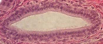
STRATIFIED (TWO-LAYER) COLUMNAR EPITHELIUM
    
Stained with haematoxylin and eosin


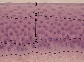
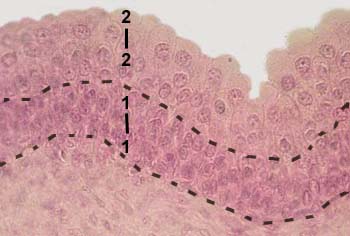
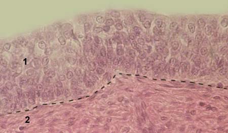
STRATIFIED SQUAMOUS NONKERATINISING EPITHELIUM
     Stained with haematoxylin and eosin

1 - basal layer
2 - intermediate layer
3 - superficial layer


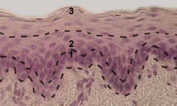
STRATIFIED SQUAMOUS NONKERATINISING EPITHELIUM
     Stained with haematoxylin and eosin

1 - basal layer
2 - intermediate layer
3 - superficial layer


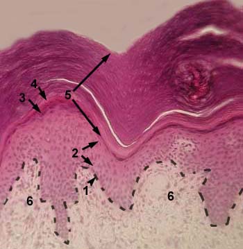
STRATIFIED SQUAMOUS KERATINISING EPITHELIUM (EPIDERMIS)
     Stained with haematoxylin and eosin

1 - basal layer
2 - prickle cell layer (stratum spinosum)
3 - granular layer
4 - lucidar layer
5 - cornified layer

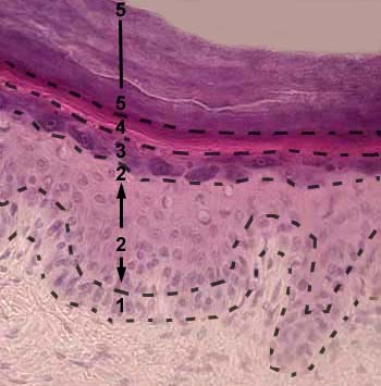
STRATIFIED SQUAMOUS KERATINISING EPITHELIUM (EPIDERMIS)
     Stained with haematoxylin and eosin

1 - basal layer
2 - prickle cell layer (stratum spinosum)
3 - granular layer
4 - lucidar layer
5 - cornified layer


TRANSITIONAL EPITHELIUM (UROTHELIUM)
     Stained with haematoxylin and eosin

1 - basal layer
2 - superficial layer


CONNECTIVE TISSUE

 
BONE

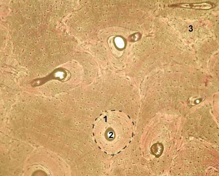
LAMELLAR (MATURE) BONE
     Stained with thionin and picric acid

1 - Haversian system (newly formed)
2 - Haversian canal
3 - interstitial system (older, partially resorbed, Haversian system)

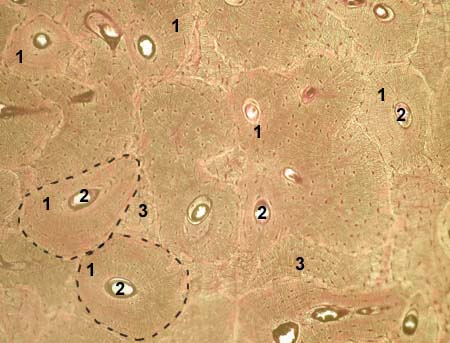
LAMELLAR (MATURE) BONE
     Stained with thionin and picric acid

1 - Haversian system (for better understanding, two Haversian systems
      are bordered with dotted line)
2 - Haversian canal
3 - interstitial system (older, partially resorbed, Haversian system)

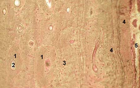
LAMELLAR (MATURE) BONE
     Stained with thionin and picric acid

1 - Haversian system (osteon)
2 - Haversian canal
3 - interstitial system (older, partially resorbed, Haversian system)
4 - outer cortical lamellae (cortical bone)
5 - periosteum

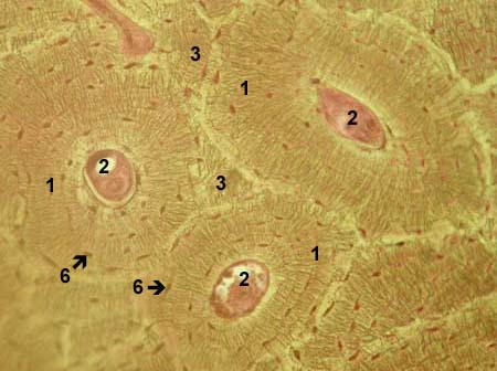
LAMELLAR (MATURE) BONE
     Stained with thionin and picric acid

1 - Haversian system (osteon)
2 - Haversian canal
3 - interstitial system (older, partially resorbed, Haversian system)
6 - osteocytes


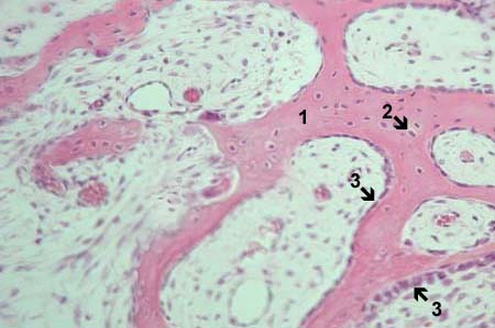
WOVEN (IMMATURE) BONE
     Stained with haematoxylin and eosin

1 - intercellular bone matrix
2 - osteocytes
3 - periosteum

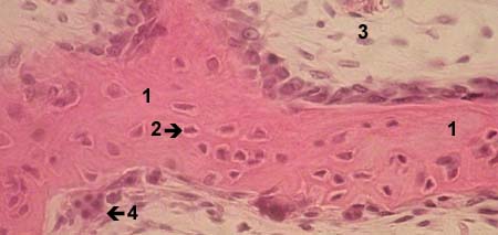
WOVEN (IMMATURE) BONE
     Stained with haematoxylin and eosin

1 - intercellular bone matrix
2 - osteocytes
3 - periosteum
4 - osteoclast

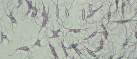
OSTEOCYTES
     Stained with haematoxylin


__________________________________________________________________


CONNECTIVE TISSUE


CARTILAGE,
DENSE FIBROUS CONNECTIVE TISSUE (TENDON)

HYALINE CARTILAGE
     Stained with haematoxylin and eosin

1 - cells of the cartilage (chondrocytes, chondroblasts)
2 - intercellular cartilage matrix
3 - perichondrium

ELASTIC CARTILAGE
     Stained with iron haematoxylin

1 - cells of the cartilage (chondrocytes, chondroblasts)
2 - intercellular cartilage matrix
3 - perichondrium


FIBROCARTILAGE (INTERVERTEBRAL DISC)
     Stained with haematoxylin and eosin

1 - cells of the cartilage (chondrocytes, chondroblasts)
2 - intercellular cartilage matrix
3 - nucleus pulposus
4 - anulus fibrosus

TENDON (longitudinal cut)
     Stained with haematoxylin and eosin

1 - endothenonium (forms I level fasciculi)
2 - perithenonium (forms II level fasciculi)

TENDON (transverse cut)
     Stained with haematoxylin and eosin

1 - endothenonium (forms I level fasciculi)
2 - perithenonium (forms II level fasciculi)


__________________________________________________________________


CONNECTIVE TISSUE


LOOSE FIBROUS CONNECTIVE TISSUE

LOOSE CONNECTIVE TISSUE
     Stained with iron haematoxylin

1 - fibroblasts
2 - collagen fibers
3 - elastin fibers

MAST CELLS
     Stained with toluidine blue

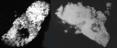
MAST CELLS
     Formaldehyde-induced fluorescence

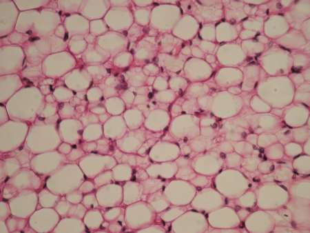
ADIPOSE

ADIPOCYTES


     Stained with sudan III       

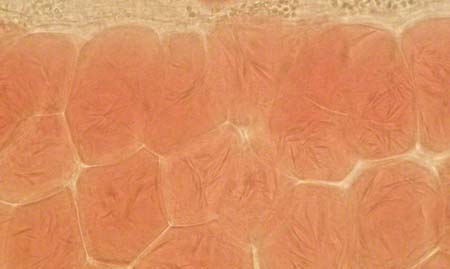
WHITE ADIPOSE TISSUE
     Stained with haematoxylin and eosin

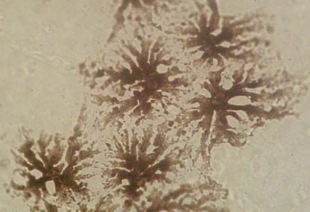
PIGMENT CELLS
     Nonstained slide


__________________________________________________________________


CONNECTIVE TISSUE

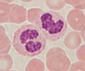
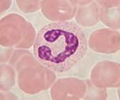
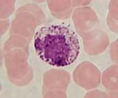
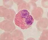
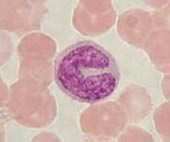
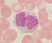
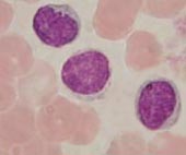
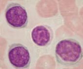
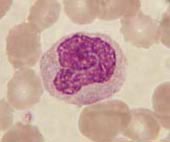
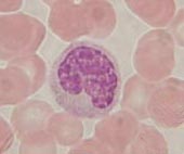
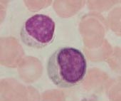
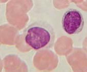
BLOOD


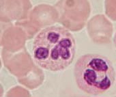
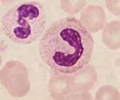
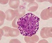
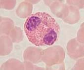
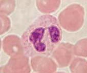
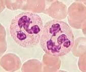
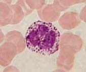
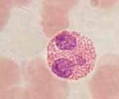
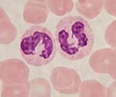
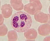
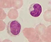
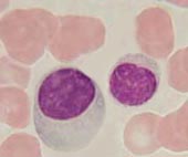
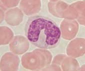
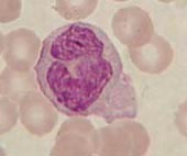
leucocytes


BASOPHILS

EOSINOPHILS

NEUTROPHILS


LYMPHOCYTES

MONOCYTES


__________________________________________________________________


CIRCULATORY SYSTEM


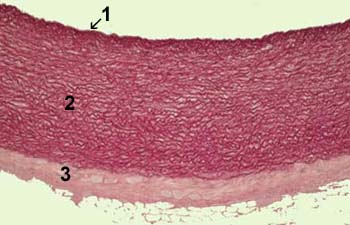
ELASTIC ARTERY (AORTA)
     Stained with orsein

1 - tunica intima
2 - tunica media
3 - tunica externa

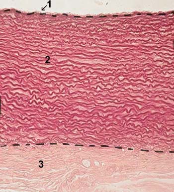
ELASTIC ARTERY (AORTA)
     Stained with orsein

1 - tunica intima
2 - tunica media
3 - tunica externa

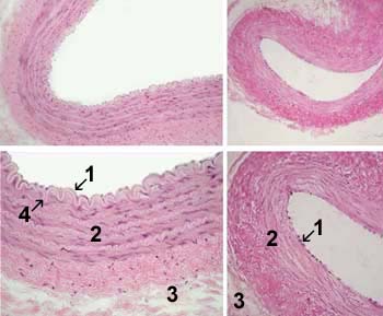
MUSCULAR ARTERY (left),
LARGE MUSCULAR VEIN (right)

     Stained with haematoxylin and eosin

1 - tunica intima
2 - tunica media
3 - tunica externa
4 - internal elastic lamina
     (is present only in arteries)

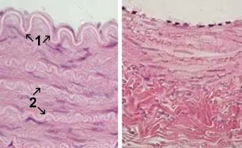
MUSCULAR ARTERY (left),
LARGE MUSCULAR VEIN (right)

     Stained with haematoxylin and eosin

1 - internal elastic lamina
     (is present only in arteries)
2 - elastic membranes within tunica media
     (are present only in arteries)

THE MICROCIRCULATION
(arterioles, venules, capillaries)

     Stained with haematoxylin and eosin

Capillaries are shown with arrows

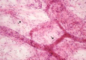
CAPILLARIES
(in the myocardium)

     Stained with haematoxylin and eosin

Capillaries are shown with arrows

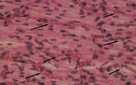
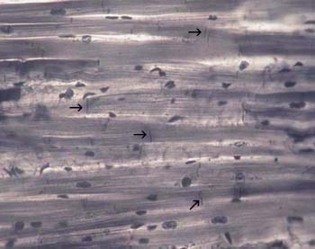
HEART (MYOCARDIUM)
     Stained with iron haematoxylin

Intercalated discs are shown with arrows


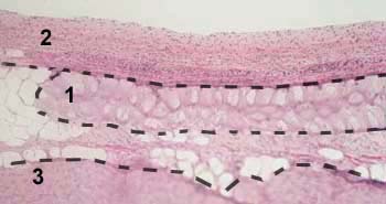
HEART (PURKINJE FIBERS)
     Stained with haematoxylin and eosin

1 - Purkinje fibers
2 - endocardium
3 - myocardium

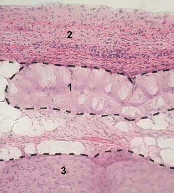
HEART (PURKINJE FIBERS)
     Stained with haematoxylin and eosin

1 - Purkinje fibers
2 - endocardium
3 - myocardium

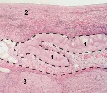
HEART (PURKINJE FIBERS)
     Stained with haematoxylin and eosin

1 - Purkinje fibers
2 - endocardium
3 - myocardium

HEART (PURKINJE FIBERS)
     Stained with haematoxylin and eosin

1 - Purkinje fibers
2 - endocardium
3 - myocardium


__________________________________________________________________


MUSCLE TISSUE

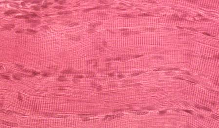
SKELETAL STRIATED (CROSS-STRIATED) MUSCLE TISSUE
longitudinal section
     Stained with haematoxylin and eosin

Longitudinally cut muscle fibers are shown;
cross-striations are seen along the fibers;
nuclei are located at peripheral parts of the fibers.

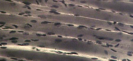
SKELETAL STRIATED MUSCLE TISSUE
longitudinal section
     Stained with iron haematoxylin

Longitudinally cut muscle fibers are shown;
cross-striations are seen along the fibers;
nuclei are located at peripheral parts of the fibers.

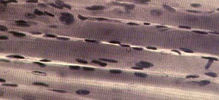
SKELETAL STRIATED MUSCLE TISSUE
longitudinal section
     Stained with iron haematoxylin

Longitudinally cut muscle fibers are shown;
cross-striations are seen along the fibers;
nuclei are located at peripheral parts of the fibers.

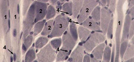
SKELETAL STRIATED MUSCLE TISSUE
transverse section
     Stained with iron haematoxylin

1 - longitudinally cut muscle fibers
2 - transversely cut muscle fibers
3 - cytoplasm of the muscle fiber (sarcoplasm)
4 - nuclei of the muscle fiber

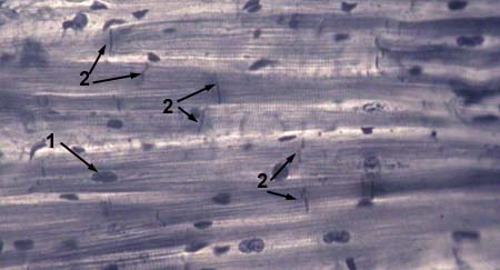
CARDIAC STRIATED MUSCLE TISSUE (MYOCARDIUM)
longitudinal section
     Stained with iron haematoxylin

1 - nuclei of cardiomyocytes
2 - intercalated discs


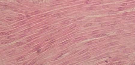
SMOOTH MUSCLE TISSUE (VISCERAL MUSCLE TISSUE)
longitudinal section
     Stained with haematoxylin and eosin

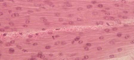
SMOOTH MUSCLE TISSUE (VISCERAL MUSCLE TISSUE)
longitudinal section
     Stained with haematoxylin and eosin


__________________________________________________________________


NERVOUS TISSUE

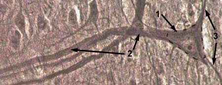
NEURONE
Impregnated with aurum chloride

1 - body of the nervous cell (pericaryon)
2 - axon
3 - dendrites

NEURONE
Impregnated with silver nitrate

1 - body of the nervous cell (pericaryon)
2 - axon
3 - dendrites

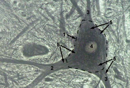
NEURONE: neurofibrills
Impregnated with silver nitrate

1 - body of the nervous cell (pericaryon)
2 - axon
3 - dendrites
4 - nucleus
5 - cytoskeleton (neuronal microtubules
      and actin filaments) is stained with
      silver as black thin filaments

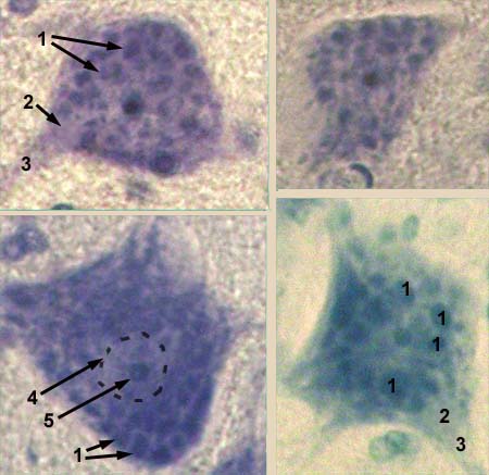
NEURONE: Nissl substance
Stained with toluidine blue by Nissl method

1 - ribosomes form clusters which are stained
      as granules or grains (Nissl substance)
2 - axon hillock
3 - axon
4 - nucleus
5 - nucleolus

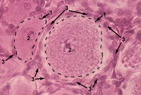
NEURONE
Stained with haematoxylin and eosin

1 - body of the nervous cell (pericaryon)
2 - nucleus of the nervous cell
3 - satellite cells which cover bodies of nervous
      cells in peripheral nervous system

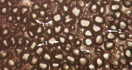
PERIPHERAL NERVE (transverse section)
Stained with osmium oxide

1 - myelinated nerve fibers (myelin sheath
      looks as dark thick ring)
3 - nonmyelinated nerve fibers

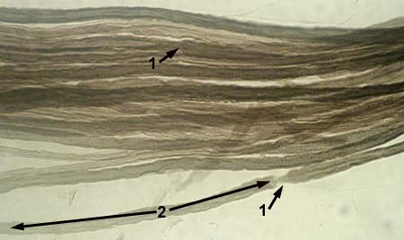
MYELINATED NERVE FIBRES
Node of Ranvier is indicated by arrow
Stained with osmium oxide

1 - node of Ranvier
2 - internode


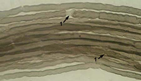
MYELINATED NERVE FIBRES
Stained with osmium oxide

1 - node of Ranvier

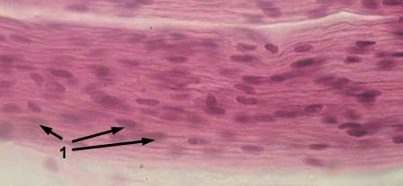
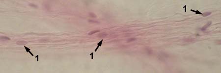
NONMYELINATED NERVE FIBRES
Stained with haematoxylin and eosin

1 - nuclei of Schwann cells

NONMYELINATED NERVE FIBRES
Stained with haematoxylin and eosin

1 - nuclei of Schwann cells

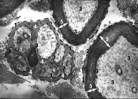
MYELINATED AND NONMYELINATED NERVE FIBRES
     Electron microscopy image

1 - nonmyelinaled nerve fibre
2 - myelinaled nerve fibre
3 - axon
4 - cytoplasm of Schwann cell, which forms nonmyelinaled sheath
5 - myelin sheath
6 - mitochondria in axon

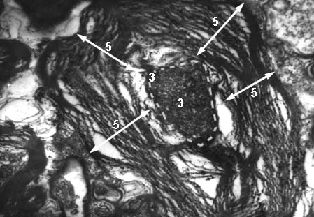
PATHOLOGICAL MYELINATED NERVE FIBRE
     Electron microscopy image

3 - wrinkled axon
5 - exfoliated myelin sheath


__________________________________________________________________


NERVOUS SYSTEM
cerebral cortex

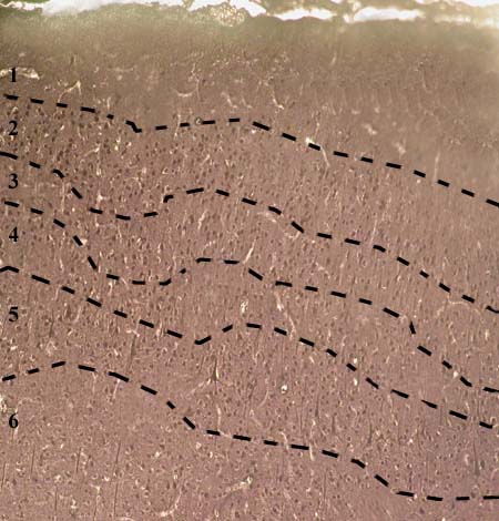
CEREBRAL CORTEX
Impregnated with silver nitrate

1 - molecular (plexiform) layer
2 - outer granular layer
3 - pyramidal cell layer
4 - inner granular layer
5 - ganglionic layer
6 - multiform cell layer

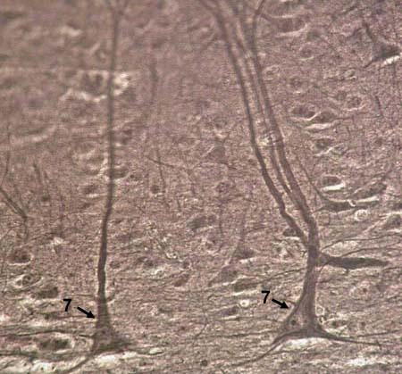
CEREBRAL CORTEX
Impregnated with silver nitrate

7 - bodies of neurons of ganglionic cell layer

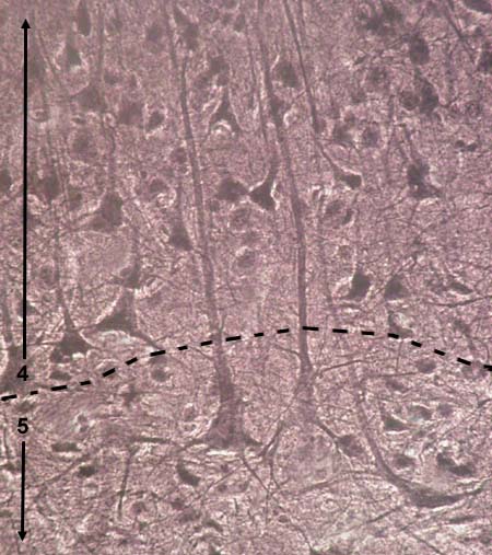
CEREBRAL CORTEX
Impregnated with silver nitrate

4 - inner granular layer
5 - ganglionic layer


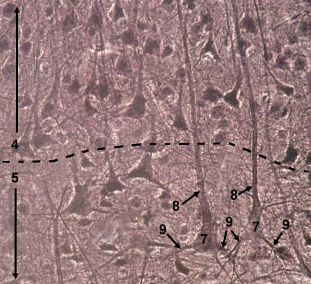
CEREBRAL CORTEX
Impregnated with silver nitrate

4 - inner granular layer
5 - ganglionic layer
7 - bodies of neurons of ganglionic cell layer
8 - àêñîíû íåéðîíîâ ãàíãëèîíàðíîãî ñëîÿ
9 - äåíäðèòû íåéðîíîâ ãàíãëèîíàðíîãî ñëîÿ

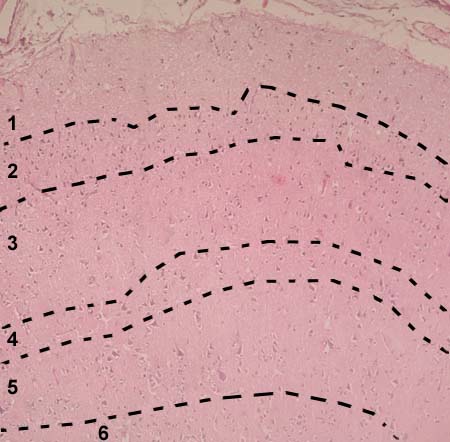
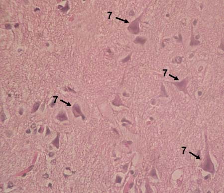
CEREBRAL CORTEX
Stained with haematoxylin and eosin

1 - molecular (plexiform) layer
2 - outer granular layer
3 - pyramidal cell layer
4 - inner granular layer
5 - ganglionic layer
6 - multiform cell layer

CEREBRAL CORTEX
Stained with haematoxylin and eosin

7 - bodies of neurons of ganglionic cell layer


__________________________________________________________________


NERVOUS SYSTEM
cerebellar cortex

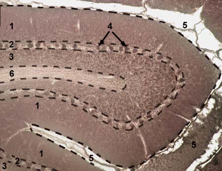
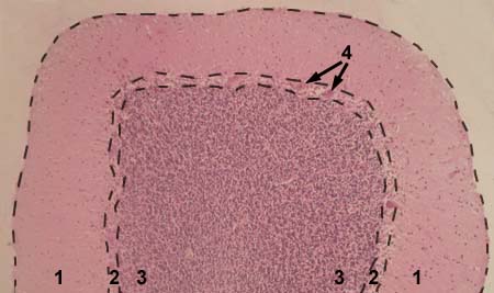
CEREBELLAR CORTEX
Impregnated with silver nitrate

1 - molecular layer
2 - ganglionic layer (Purkinje cell layer)
3 - granular cell layer
4 - Purkinje neurons (cells)
5 - fold of the cortex
6 - white matter

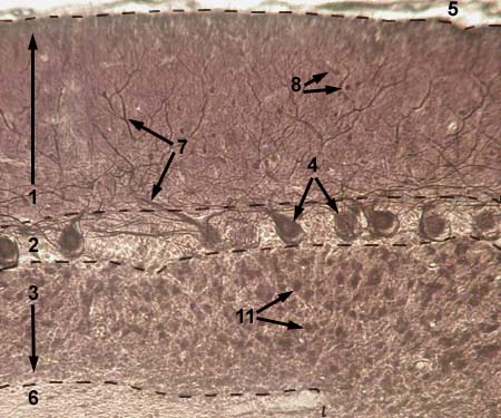
CEREBELLAR CORTEX
Impregnated with silver nitrate

1 - molecular layer
2 - ganglionic layer (Purkinje cell layer)
3 - granular cell layer
4 - Purkinje neurons (cells)
5 - fold of the cortex
6 - white matter
7 - dendrites of Purkinje cells
8 - stellate neurons (cells) and basket neurons (cells)
11 - granular neurons (cells)

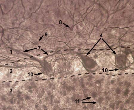
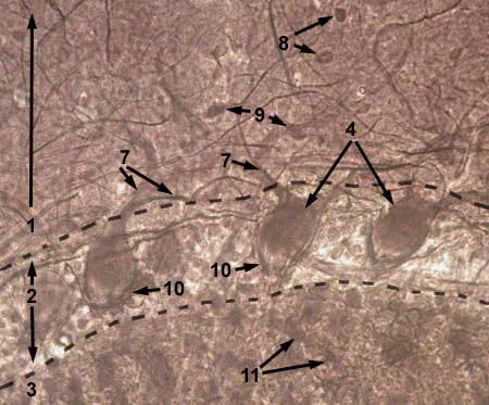
CEREBELLAR CORTEX
Impregnated with silver nitrate

1 - molecular layer
2 - ganglionic layer (Purkinje cell layer)
3 - granular cell layer
4 - Purkinje neurons (cells)
5 - fold of the cortex
6 - white matter
7 - dendrites of Purkinje cells
8 - stellate neurons (cells) and basket neurons (cells)
9 - stellate neurons
10 - axons of stellate and basket neurons, 
     twining cell bodies of Purkinje neurons
11 - granular neurons (cells)

CEREBELLAR CORTEX
Impregnated with silver nitrate

1 - molecular layer
2 - ganglionic layer (Purkinje cell layer)
3 - granular cell layer
4 - Purkinje neurons (cells)
7 - dendrites of Purkinje cells
8 - stellate neurons (cells) and basket neurons (cells)
9 - stellate neurons
10 - axons of stellate and basket neurons, 
     twining cell bodies of Purkinje neurons
11 - granular neurons (cells)


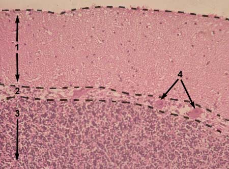
CEREBELLAR CORTEX
Stained with haematoxylin and eosin

1 - molecular layer
2 - ganglionic layer (Purkinje cell layer)
3 - granular cell layer
4 - Purkinje neurons (cells)

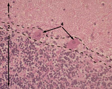
CEREBELLAR CORTEX
Stained with haematoxylin and eosin

1 - molecular layer
2 - ganglionic layer (Purkinje cell layer)
3 - granular cell layer
4 - Purkinje neurons (cells)

CEREBELLAR CORTEX
Stained with haematoxylin and eosin

1 - molecular layer
2 - ganglionic layer (Purkinje cell layer)
3 - granular cell layer
4 - Purkinje neurons (cells)


__________________________________________________________________


NERVOUS SYSTEM
spinal cord, ganglia

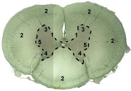
SPINAL CORD
Impregnated with silver nitrate

1 - gray matter
2 - white matter
3 - ventral horns of gray matter
4 - dorsal horns of gray matter
5 - lateral horns of gray matter

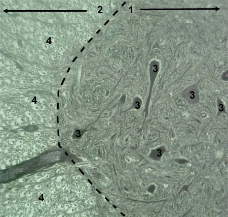
SPINAL CORD
Impregnated with silver nitrate

1 - gray matter (ventral horns)
2 - white matter
3 - nervous cell bodies 
4 - nervous fibers in transverse cut 
      (convey tracts)

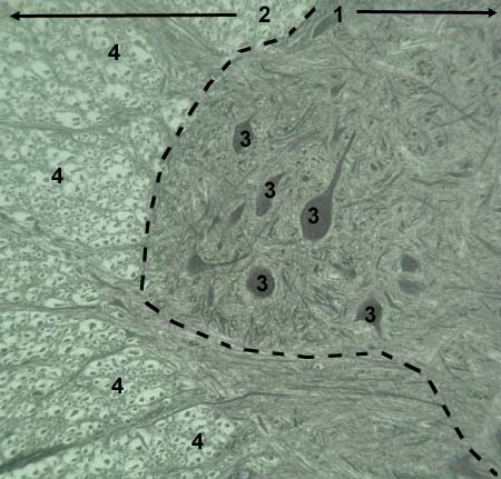
SPINAL CORD
Impregnated with silver nitrate

1 - gray matter (ventral horns)
2 - white matter
3 - nervous cell bodies 
4 - nervous fibers in transverse cut 
      (convey tracts)

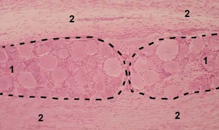
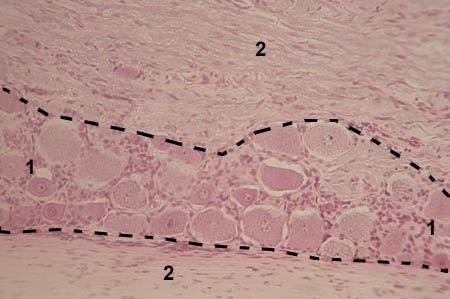
SPINAL GANGLION
Stained with haematoxylin and eosin

1 - cell bodies of neurons (located by groups)
2 - nerve fibers

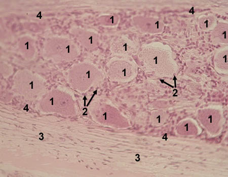
SPINAL GANGLION
Stained with haematoxylin and eosin

1 - cell bodies of neurons (pseudo-unipolar neurons)
2 - nerve fibers

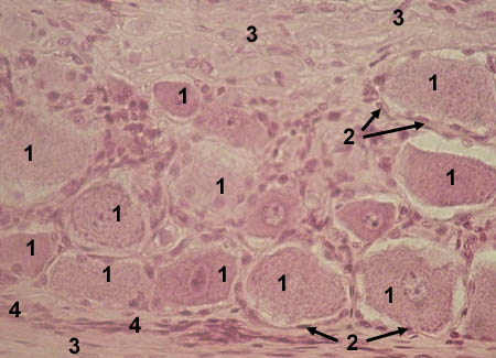
SPINAL GANGLION
Stained with haematoxylin and eosin

1 - cell bodies of neurons (pseudo-unipolar neurons)
2 - satellite cells
3 - nerve fibers
4 - interlayers of connective tissue

SPINAL GANGLION
Stained with haematoxylin and eosin

1 - cell bodies of neurons (pseudo-unipolar neurons)
2 - satellite cells
3 - nerve fibers
4 - interlayers of connective tissue


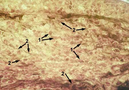
SYMPATHETIC GANGLION
Impregnated with silver nitrate

1 - cell bodies of neurons (multipolar neurons)
2 - nerve fibers

SYMPATHETIC GANGLION
Impregnated with silver nitrate

1 - cell bodies of neurons (multipolar neurons)
2 - nerve fibers


__________________________________________________________________


SENSE ORGANS

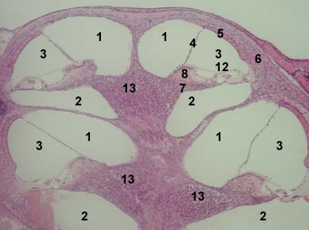
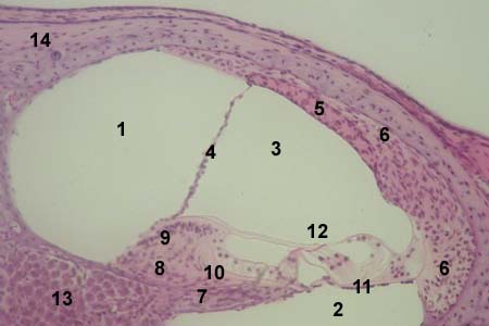
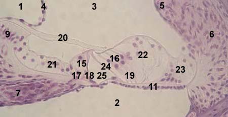
ORGAN OF CORTI (organ of hear)
     Stained with haematoxylin and eosin

1 - scala vestibuli
2 - scala media of the cochlear
3 - scala tympani
4 - vestibular membrane
5 - stria vascularis
6 - spiral ligament
7 - osseous spiral lamina
8 - spiral limbus (thickened mass above the free
    edge of the spiral lamina)
9 - vestibular surface (lip) of the limbus
10 - tympanic surface (lip) of the limbus
11 - basilar membrane
12 - organ of Corti
13 - spiral ganglion
14 - osseous labyrinth of the cochlear


15 - inner sensory (hair) cells
16 - outer sensory (hair) cells
17 - inner phalangeal cells
18 - inner pillar cells
19 - outer phalangeal cells
20 - tectorial membrane
21 - inner supporting cells
22 - intermediate supporting cells
23 - border supporting cells
24 - outer pillar cells
25 - tunnel of Corti

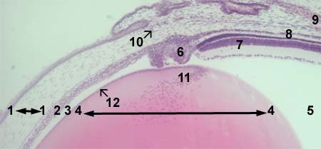
EYE
     Stained with haematoxylin and eosin

1 - cornea
2 - iris
3 - posterior chamber of the eye
4 - lens
5 - vitreous body
6 - ciliary body
7 - retina
8 - choroid
9 - sclera
10 - canal of Sclemm
11 - growth area of the lens
12 - anterior epithelium of the lens

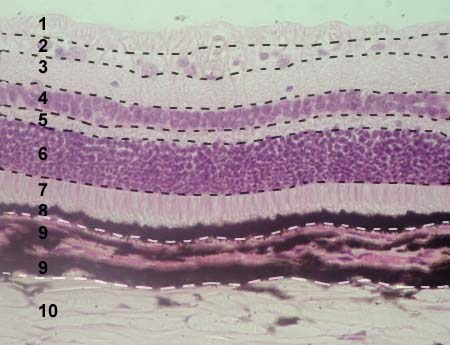
EYE: RETINA, CHOROID
     Stained with haematoxylin and eosin

1 - 8 - retina
1 - optic nerve fibers
2 - ganglion cell layer
3 - inner plexiform layer
4 - inner nuclear layer
5 - outer plexiform layer
6 - outer nuclear layer
7 - outer processes of rods and cones
8 - pigmented epithelium
9 - choroid
10 - sclera

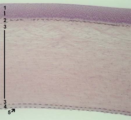
EYE: CORNEA
     Stained with haematoxylin and eosin

1 - anterior epithelium (stratified
      squamous epithelium)
2 - anterior basement (Bowman's) membrane
3 - substantia propria
4 - posterior basement (Descemet's) membrane
5 - posterior epithelium (simple squamous or endothelium)

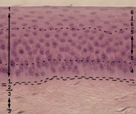
EYE: CORNEA
     Stained with haematoxylin and eosin anterior epithelium (stratified
      squamous epithelium)
2 - anterior basement (Bowman's) membrane
3 - substantia propria
4 - basal layer of the epithelium
5 - intermediate layer of the epithelium
6 - superficial layer of the epithelium


EYE: IRIS
     Stained with haematoxylin and eosin

1 - anterior epithelium
2 - anterior terminal layer
3 - vascular layer
4 - posterior terminal layer
5 - pigmented layer
6 - blood vessel
7 - dilator pupillae musle
8 - constrictor pupillae musle

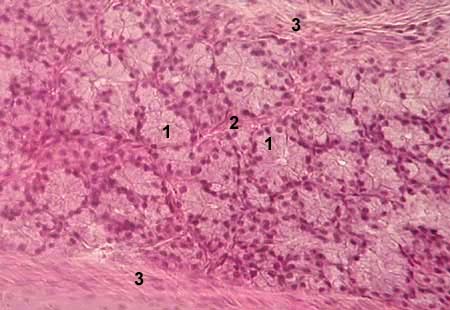
LACRIMAL GLAND
     Stained with haematoxylin and eosin

1 - secretory (end) unit
2 - interlobular connective tissue
3 - capsule

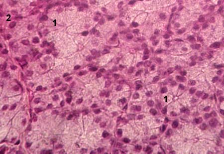
LACRIMAL GLAND
     Stained with haematoxylin and eosin

1 - secretory (end) unit
2 - intralobular excretory duct

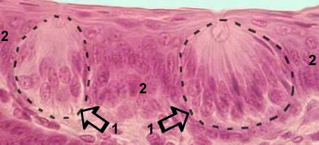
TASTE BUDS
     Stained with haematoxylin and eosin

1 - taste buds
2 - stratified squamous epithelium of
     the mucosa of the tongue

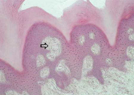
MEISSNER'S CORPUSCLE
in the dermis of the skin of the fingertip
     Stained with haematoxylin and eosin

     Meissner's corpuscle (oval in shape)
     showed with arrow

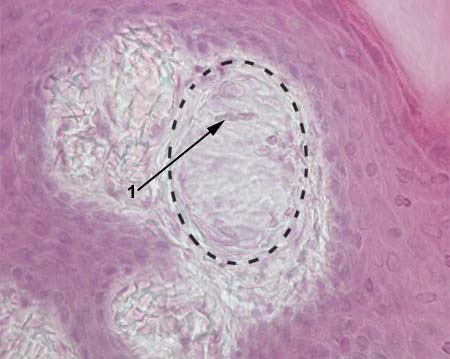
MEISSNER'S CORPUSCLE
in the skin of the fingertip
     Stained with haematoxylin and eosin

     Meissner's corpuscle encircled with dotted line

1 - arrow shows nuclei of modified 
     neuroglial cells

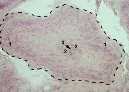
PACINIAN CORPUSCLE
     Stained with haematoxylin and eosin

          Pacinian corpuscle encircled
          with dotted line
1 - multilayered connective tissue capsule
2 - modified neuroglial cells
      (form inned calsule)
3 - dendrite of a sensory neuron

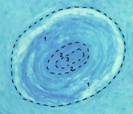
HERBST AND GRANDRY SENSORY CORPUSCLE
     Stained with haematoxylin and eosin

          Herbst and Grandry corpuslcle encircled
          with dotted line
1 - multilayered connective tissue capsule
2 - modified neuroglial cells
      (form inned calsule or cone)
3 - dendrite of a sensory neuron


__________________________________________________________________


DIGESTIVE SYSTEM


ESOPHAGUS, STOMACH


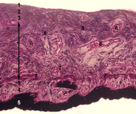
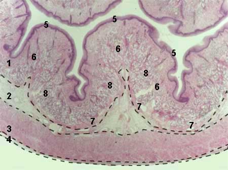
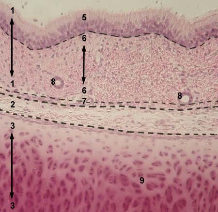
ESOPHAGUS
     Stained with haematoxylin and eosin

1 - tunica mucosa
2 - tunica submucosa
3 - tunica muscularis propria
4 - tunica adventitia
5 - epithelium of the mucosa
6 - lamina propria of the mucosa
7 - muscularis mucosae
8 - glands in the lamina propria

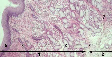
ESOPHAGUS
     Stained with haematoxylin and eosin

1 - tunica mucosa
2 - tunica submucosa
5 - epithelium of the mucosa
6 - lamina propria of the mucosa
7 - muscularis mucosae
8 - glands in the lamina propria

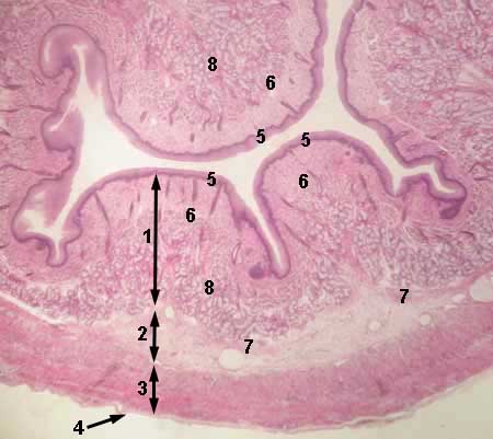
ESOPHAGUS
     Stained with haematoxylin and eosin

1 - tunica mucosa
2 - tunica submucosa
3 - tunica muscularis propria
4 - tunica adventitia
5 - epithelium of the mucosa
6 - lamina propria of the mucosa
7 - muscularis mucosae
8 - glands in the lamina propria

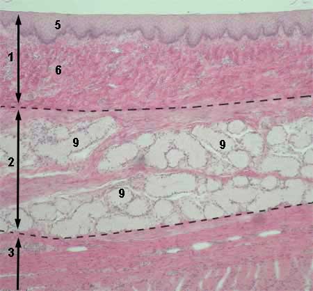
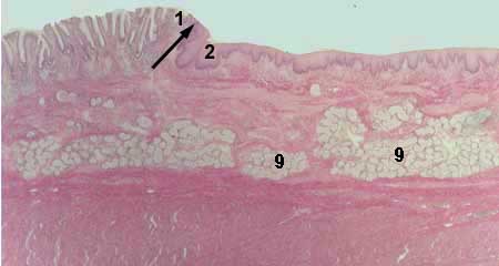
ESOPHAGUS
     Stained with haematoxylin and eosin

1 - tunica mucosa
2 - tunica submucosa
3 - tunica muscularis propria
5 - epithelium of the mucosa
6 - lamina propria of the mucosa
9 - glands in the submucosa

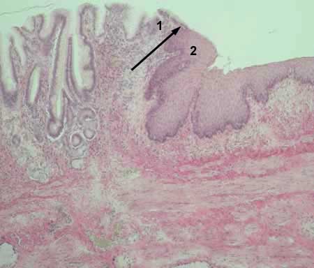
GASTRO-ESOPHAGEAL JUNCTION
     Stained with haematoxylin and eosin

1 - stomach
2 - esophagus

GASTRO-ESOPHAGEAL JUNCTION
     Stained with haematoxylin and eosin

1 - stomach
2 - esophagus


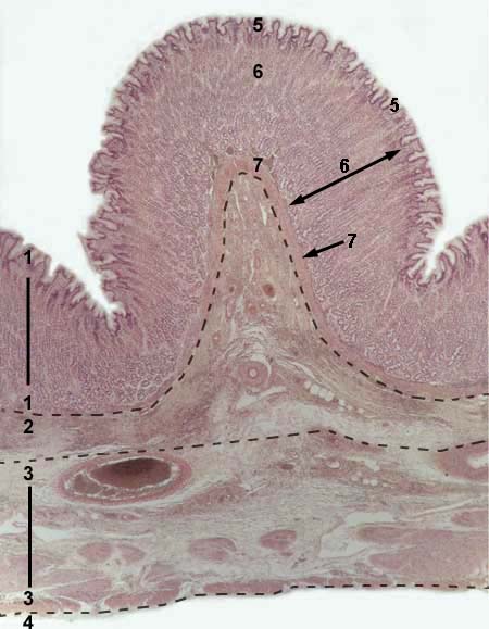
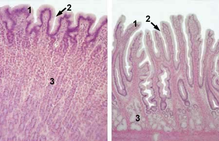
FUNDAL PART OF THE STOMACH
     Stained with haematoxylin and eosin

1 - tunica mucosa
2 - tunica submucosa
3 - tunica muscularis propria
4 - tunica serosa
5 - epithelium of the mucosa
6 - lamina propria of the mucosa (contains glands)
7 - muscularis mucosae

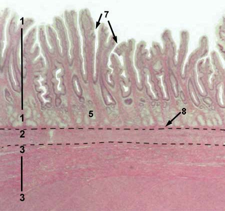
PYLORIC PART OF THE STOMACH
     Stained with haematoxylin and eosin

1 - tunica mucosa
2 - tunica submucosa
3 - tunica muscularis propria
5 - lamina propria of the mucosa     
     (contains glands)
7 - gastric pits in the mucosa
8 - muscularis mucosae

FUNDAL PART (left),
PYLORIC PART (right)
OF THE STOMACH

     Stained with haematoxylin and eosin

1 - epithelium of the mucosa
2 - gastric pits in the mucosa
3 - glands in the lamina propria


__________________________________________________________________


DIGESTIVE SYSTEM

 
INTESTINE


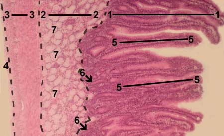
DUODENUM
     Stained with haematoxylin and eosin

1 - tunica mucosa
2 - tunica submucosa
3 - tunica muscularis propria
4 - tunica serosa
5 - villi
6 - glands (crypts) in the lamina
      propria of the mucosa
7 - glands in the tunica submucosa (Brunner's glands)

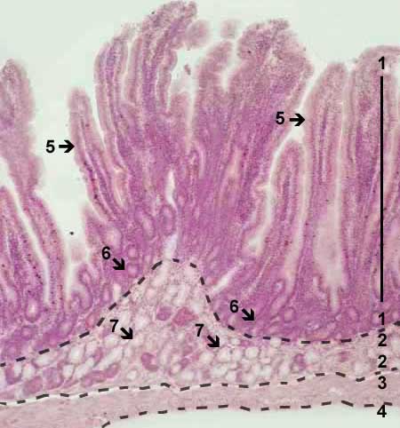
DUODENUM
     Stained with haematoxylin and eosin

1 - tunica mucosa
2 - tunica submucosa
3 - tunica muscularis propria
4 - tunica serosa
5 - villi
6 - glands (crypts) in the lamina
     propria of the mucosa
7 - glands in the tunica submucosa (Brunner's glands)

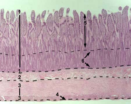
JEJUNUM
     Stained with haematoxylin and eosin

1 - tunica mucosa
2 - tunica submucosa
3 - tunica muscularis propria
4 - tunica serosa
5 - villi
6 - glands (crypts) in the lamina
     propria of the mucosa

JEJUNUM
     Stained with haematoxylin and eosin

1 - epithelium of the mucosa
      (covers villi)
2 - lamina propria of the mucosa
3 - goblet cells in the epithelium
4 - parts of the muscularis mucosae

JEJUNUM
     Stained with haematoxylin and eosin

1 - epithelium of the mucosa
      (covers villi)
2 - lamina propria of the mucosa
3 - goblet cells in the epithelium
4 - parts of the muscularis mucosae


ILEUM
     Stained with haematoxylin and eosin

1 - tunica mucosa
2 - tunica submucosa
3 - tunica muscularis propria
4 - tunica serosa
5 - villi
6 - epithelium of the mucosa
      (covers villi)
7 - connective tissue of the lamina
      propria of the mucosa
6 - glands (crypts) in the lamina
      propria of the mucosa

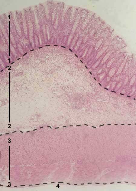
COLON
     Stained with haematoxylin and eosin

1 - tunica mucosa
2 - tunica submucosa
3 - tunica muscularis propria
4 - tunica serosa
5 - lymphoid follicle in the lamina
     propria of the mucosa

COLON
     Stained with haematoxylin and eosin

1 - tunica mucosa
2 - tunica submucosa
3 - tunica muscularis propria
4 - tunica serosa

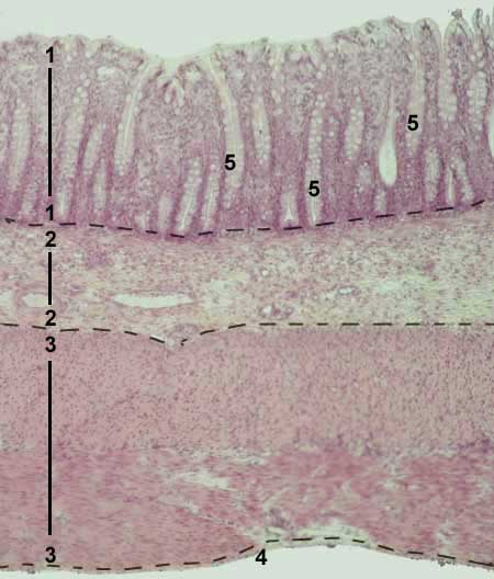
COLON
     Stained with haematoxylin and eosin

1 - tunica mucosa
2 - tunica submucosa
3 - tunica muscularis propria
4 - tunica serosa
5 - glands (crypts) in the lamina
     propria of the mucosa

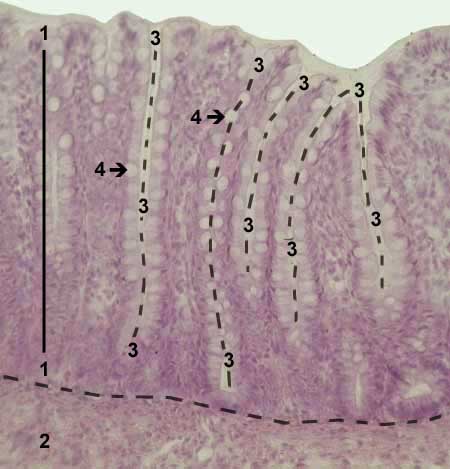
COLON
     Stained with haematoxylin and eosin

1 - tunica mucosa
2 - tunica submucosa
3 - glands (crypts) in the lamina
     propria of the mucosa
4 - goblet cells in the epithelium of crypts

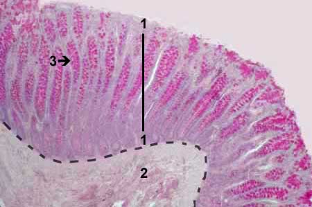
COLON
     Stained with mucicarmin

1 - tunica mucosa
2 - tunica submucosa
3 - goblet cells in the epithelium of crypts
      (stained with red-magenta color)


__________________________________________________________________


DIGESTIVE SYSTEM

 
LIVER, PANCREAS

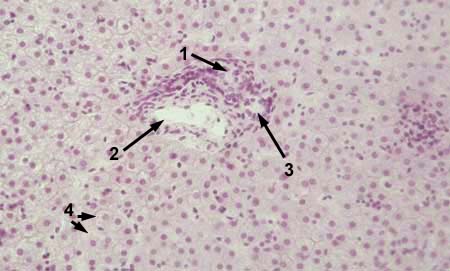
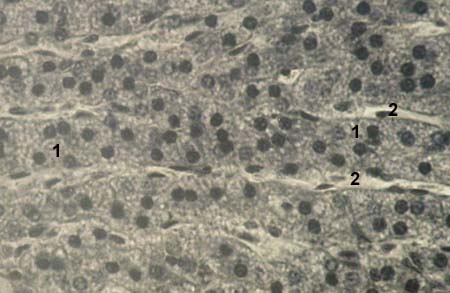
LIVER
     Stained with haematoxylin and eosin

1 - hepatic artery
2 - portal vien
3 - bile duct
4 - hepatocytes
1, 2, 3 - portal tract

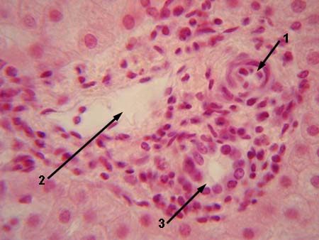
LIVER
     Stained with haematoxylin and eosin

1 - hepatic artery
2 - portal vien
3 - bile duct
4 - hepatocytes
1, 2, 3 - portal tract

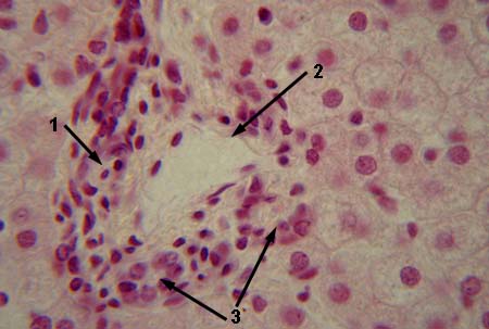
LIVER
     Stained with haematoxylin and eosin

1 - hepatic artery
2 - portal vien
3 - bile duct
1, 2, 3 - portal tract

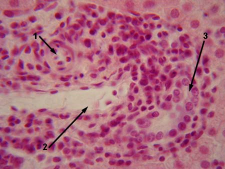
LIVER
     Stained with haematoxylin and eosin

1 - hepatic artery
2 - portal vien
3 - bile duct
1, 2, 3 - portal tract

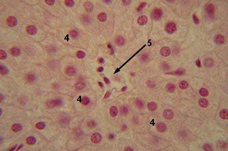
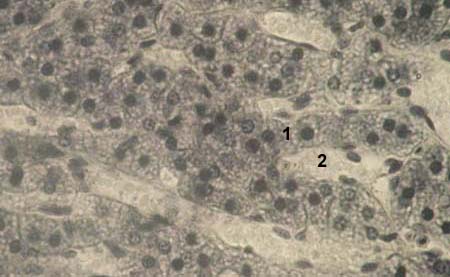
LIVER
     Stained with haematoxylin and eosin

4 - hepatocytes
5 - terminal hepatic (centrilobular) venule

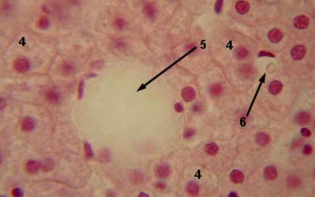
LIVER
     Stained with haematoxylin and eosin

4 - hepatocytes
5 - terminal hepatic (centrilobular) venule
6 - hepatic sinusoid

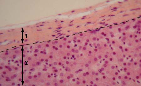
LIVER
     Stained with haematoxylin and eosin

1 - capsule
2 - liver parenchyma

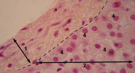
LIVER
     Stained with haematoxylin and eosin

1 - capsule
2 - liver parenchyma
4 - hepatocytes


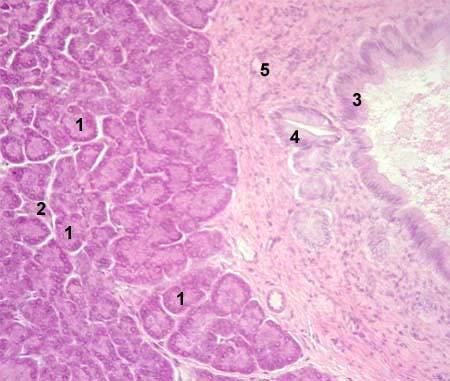
PANCREAS
     Stained with haematoxylin and eosin

1 - glandular acinus
2 - islet of Langerhans
3 - main duct
4 - interlobular duct
5 - interlobular connective tissue septa

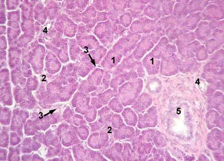
PANCREAS
     Stained with haematoxylin and eosin

1 - acinus
2 - islet of Langerhans
3 - interlobular connective tissue septa
4 - intralobular duct
5 - interlobular duct

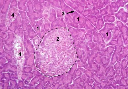
PANCREAS
     Stained with haematoxylin and eosin

1 - acinus
2 - islet of Langerhans
3 - interlobular connective tissue septa
4 - blood vessels

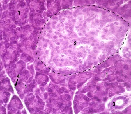
PANCREAS
     Stained with haematoxylin and eosin

1 - acinus
2 - islet of Langerhans
3 - intralobular duct
4 - interlobular connective tissue septa


________________________________________________________


DIGESTIVE SYSTEM

 
TONGUE, TOOTH, TOOTH DEVELOPMENT


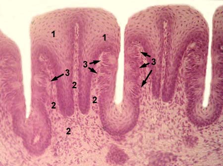
FILIFORM PAPILLAE OF THE TONGUE
     Stained with haematoxylin and eosin

1 - epithelium covering papilla
      (stratified squamous keratinizing)
2 - keratinized layer of the epithelium
3 - core of the papilla (lamina propria
      of the mucosa of dorsal surface of the tongue)
2 - tongue muscles

LEAF SHAPED PAPILLAE OF THE TONGUE
     Stained with haematoxylin and eosin

1 - epithelium covering papilla
      (stratified squamous nonkeratinizing)
2 - core of the papilla (lamina propria
      of the mucosa of dorsal surface of the tongue)
3 - taste buds

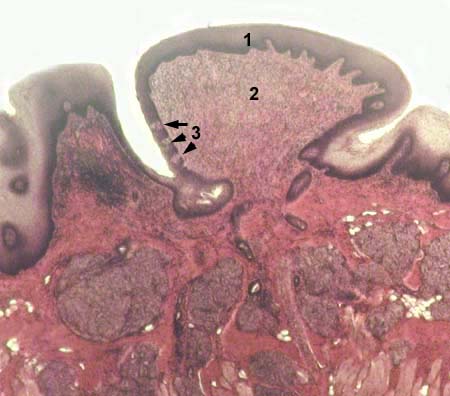
CIRCUMVALLATE PAPILLAE OF THE TONGUE
     Stained with haematoxylin and eosin

1 - epithelium covering papilla
      (stratified squamous nonkeratinizing)
2 - core of the papilla (lamina propria
      of the mucosa of dorsal surface of the tongue)
3 - taste buds

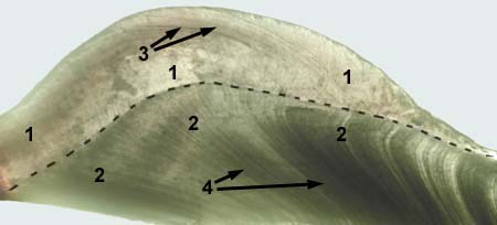
TOOTH
     Nonstained

1 - enamel
2 - dentine
3 - Retcius lines
4 - dentine tubules


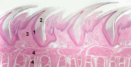
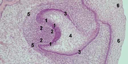
TOOTH DEVELOPMENT - ENAMEL ORGAN
     Stained with haematoxylin and eosin

1, 3, 4 - enamel organ
1 - internal cells of the enamel organ
      (these cells will be ameloblsats)
2 - dental papilla
3 - external cells of the enamel organ
4 - cells forming the main bulk of
      the enamel organ (stellate reticulum)
5 - dental follicle
6 - epithelium of oral cavity

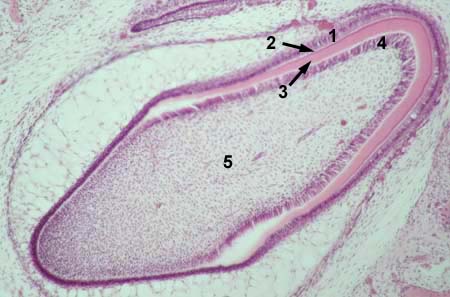
TOOTH DEVELOPMENT - FORMATION OF DENTAL TISSUES
     Stained with haematoxylin and eosin

1 - ameloblasts (former external cells of the enamel organ)
2 - enamel
3 - dentine (predentine)
4 - odontoblasts (cells which covered the top of dental papilla)
5 - dental pulp (former dental papilla)

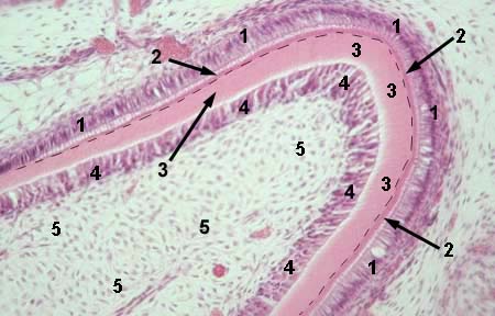
TOOTH DEVELOPMENT - FORMATION OF DENTAL TISSUES
     Stained with haematoxylin and eosin

1 - ameloblasts
2 - enamel
3 - dentine (predentine)
4 - odontoblasts
5 - dental pulp
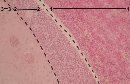
  border between enamel and dentine
  is marked with dot line


__________________________________________________________________


 ENDOCRINE SYSTEM

 
PITUITARY GLAND (HYPOPHYSIS), PINEAL GLAND (EPIPHYSIS)


MAMMALIAN PITUITARY GLAND
     Stained with haematoxylin and eosin

1 - anterior piruitary
2 - pars intermediata
3 - pars posterior

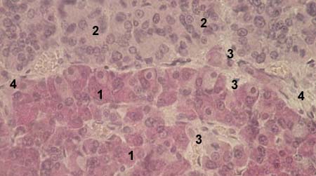
HUMAN PITUITARY GLAND,
ANTERIOR PITUITARY, ADENOHYPOPHYSIS
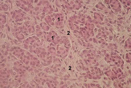
     Stained with haematoxylin and eosin

1 - acidophil cells
2 - basophil cells
3 - chromophobe cells
4 - thin streaks of connective tissue

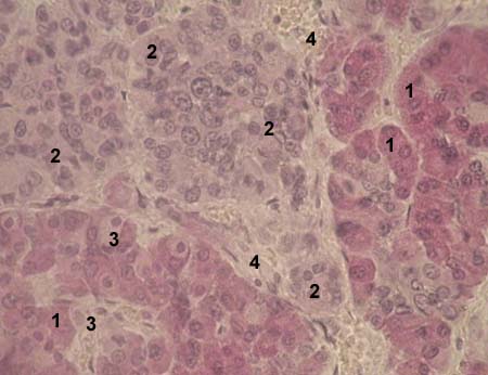
HUMAN PITUITARY GLAND,
ANTERIOR PITUITARY, ADENOHYPOPHYSIS
     Stained with haematoxylin and eosin

1 - acidophil cells
2 - basophil cells
3 - chromophobe cells
4 - thin streaks of connective tissue

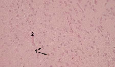
HUMAN PITUITARY GLAND, PARS POSTERIOR
     Stained with haematoxylin and eosin

1 - nuclei of pituicytes
2 - blood vessels

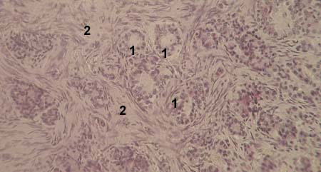
HUMAN PITUITARY GLAND,
ANALOGUE OF PARS INTERMEDIATA

     Stained with haematoxylin and eosin

1 - follicle-like structures
2 - thin streaks of connective tissue


EPIPHYSIS (PINEAL GLAND)
     Stained with haematoxylin and eosin

1 - pinealocytes
2 - pineal sand (salts of calcium, magnesium and
      silicon

EPIPHYSIS (PINEAL GLAND)
     Stained with haematoxylin and eosin

1 - pinealocytes
2 - pineal sand (salts of calcium, magnesium and
      silicon
3 - blood vessels


__________________________________________________________________


ENDOCRINE SYSTEM

 
THYROID GLAND, PARATHYROID LAND, ADRENAL GLAND


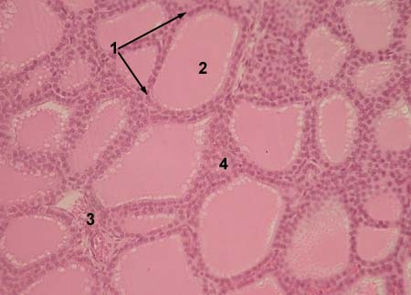
THYROID GLAND
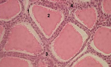
     Stained with haematoxylin and eosin

1 - thyroid follicle
2 - cavity of the thyroid follicle, filled with colloid
3 - blood vessel
4 - parafollicular cells forming interfollicular clumps

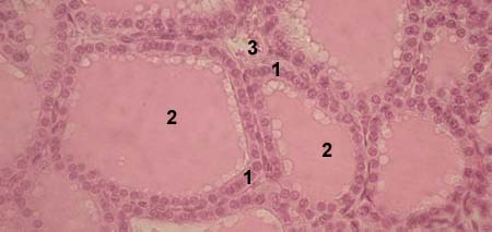
THYROID GLAND
     Stained with haematoxylin and eosin

1 - wall of the thyroid follicle, built from thyrocytes
2 - cavity of the thyroid follicle, filled with colloid
3 - blood vessel

THYROID GLAND
     Stained with haematoxylin and eosin

1 - wall of the thyroid follicle, built from thyrocytes
2 - cavity of the thyroid follicle, filled with colloid
3 - blood vessel
4 - parafollicular cells (C-cells)

PARATHYROID GLAND
     Stained with haematoxylin and eosin

1 - parathyrocytes
2 - thin streaks of connective tissue


ADRENAL GLAND (SUPRARENAL GLAND)
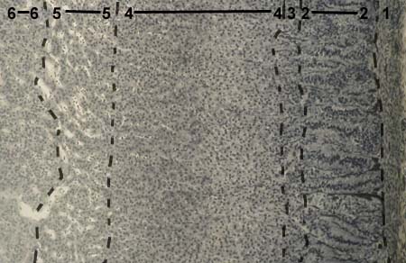
     Stained with haematoxylin and eosin

1 - outer capsule of the adrenal gland
2 - zona glomerulosa of the cortex
3 - sudanophobe layer of the cortex
4 - zona fasciculata of the cortexà
5 - zona reticularis of the cortex
6 - medulla


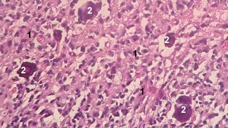
ADRENAL GLAND (SUPRARENAL GLAND)
zona glomerulosa of the cortex
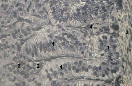
     Stained with iron haematoxylin

1 - endocrine cells of the zona glomerulosa
2 - sudanophobe layer, contained
      low differentiated cells
3 - capsule of the adrenal gland
4 - wide diameter capillares


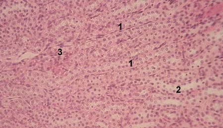
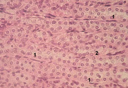
ADRENAL GLAND (SUPRARENAL GLAND)
zona fasciculata of the cortex
     Stained with iron haematoxylin

1 - endocrine cells of the zona fasciculata
2 - wide diameter capillares


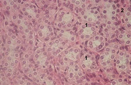
ADRENAL GLAND (SUPRARENAL GLAND)
zona reticularis of the cortex
     Stained with iron haematoxylin

1 - endocrine cells of the zona reticularis
2 - wide diameter capillares


__________________________________________________________________


RESPIRATORY SYSTEM


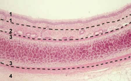
TRACHEA
     Stained with haematoxylin and eosin

1 - tunica mucosa
2 - tunica submucosa
3 - fibro-elastic layer
4 - tunica adventitia

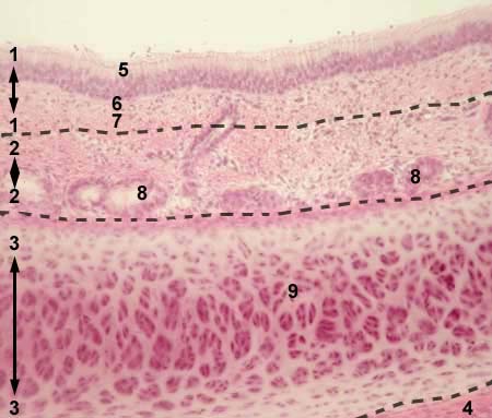
TRACHEA
     Stained with haematoxylin and eosin

1 - tunica mucosa
2 - tunica submucosa
3 - fibro-elastic layer
4 - tunica adventitia
5 - epithelium of the mucosa
6 - lamina propria of the mucosa
7 - lamina muscularis of the mucosa
8 - glands in the tunica submucosa
9 - hyaline cartilage

TRACHEA
     Stained with haematoxylin and eosin

1 - tunica mucosa
2 - tunica submucosa
3 - fibro-elastic layer
5 - epithelium of the mucosa
6 - lamina propria of the mucosa
7 - lamina muscularis of the mucosa
8 - evacuatory ducts of submucosal glands
9 - hyaline cartilage

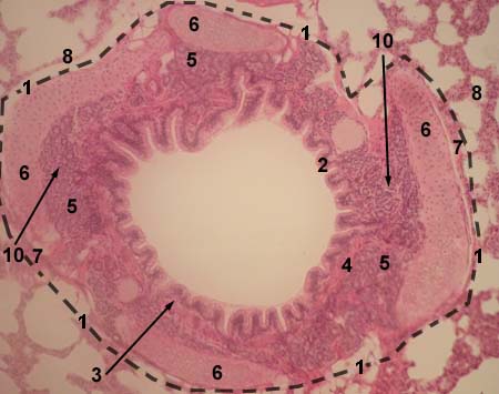
LUNG
     Stained with haematoxylin and eosin

1 - middle diameter bronchus
2 - epithelium of the mucosa
3 - lamina propria of the mucosa
4 - lamina muscularis of the mucosa
5 - tunica submucosa
6 - fibro-elastic layer
7 - tunica adventitia
8 - alveoli
9 - interstitial connective tissue of the lung
10 - glands in tunica submucosa

NOTE!
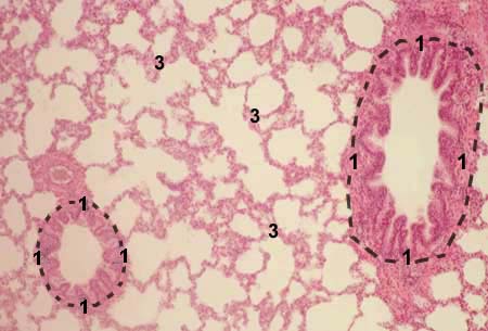
There is bronchial system in the lung, from main, or primary, bronchi to bronchioles.
Morphologically, there are only two types of bronches in the lung.

  • One type is large and middle diameter bronchi. This type of bronchi have tunica submucosa and fibro-elastic layer.
  • Another type of bronchi is small diameter bronchi. It includes small bronchi and bronchioles. This type has not both submucosal fibro-elastic layers.

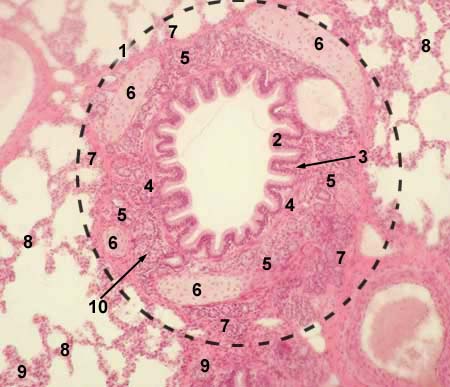
LUNG
     Stained with haematoxylin and eosin

1 - middle diameter bronchus
2 - epithelium of the mucosa
3 - lamina propria of the mucosa
4 - lamina muscularis of the mucosa
5 - tunica submucosa
6 - fibro-elastic layer
7 - tunica adventitia
8 - alveoli
9 - interstitial connective tissue of the lung
10 - glands in tunica submucosa


LUNG
     Stained with haematoxylin and eosin

1 - small diameter bronchus
3 - alveoli

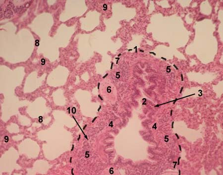
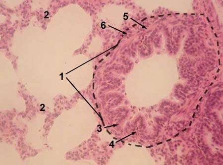
LUNG
     Stained with haematoxylin and eosin

1 - small diameter bronchus
2 - alveoli
3 - epithelium of the mucosa
4 - lamina propria of the mucosa
5 - tunica muscularis propria
6 - tunica adventitia

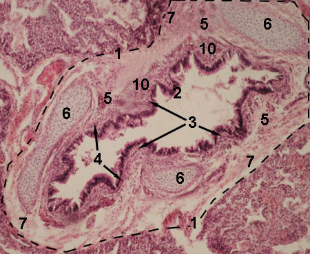
LUNG
     Stained with haematoxylin and eosin

1 - middle diameter bronchus
2 - epithelium of the mucosa
3 - lamina propria of the mucosa
4 - lamina muscularis of the mucosa
5 - tunica submucosa
6 - fibro-elastic layer
7 - tunica adventitia
10 - glands in tunica submucosa

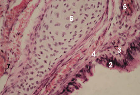
LUNG
middle diameter bronchus

     Stained with haematoxylin and eosin

2 - epithelium of the mucosa
3 - lamina propria of the mucosa
4 - lamina muscularis of the mucosa
5 - tunica submucosa
6 - fibro-elastic layer
7 - tunica adventitia


__________________________________________________________________


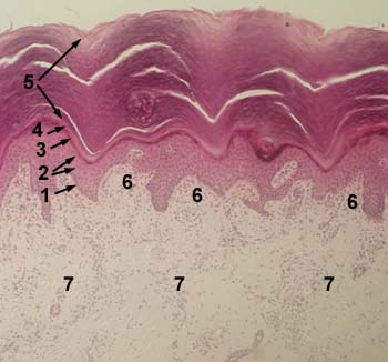
SKIN


SKIN OF THE FINGER
Stained with haematoxylin and eosin

1 - basal layer of epidermis
2 - prickle cell layer (stratum spinosum) of epidermis
3 - granular layer of epidermis
4 - lucidar layer of epidermis
5 - cornified layer of epidermis
6 - papillary layer of dermis
7 - reticular layer of dremis

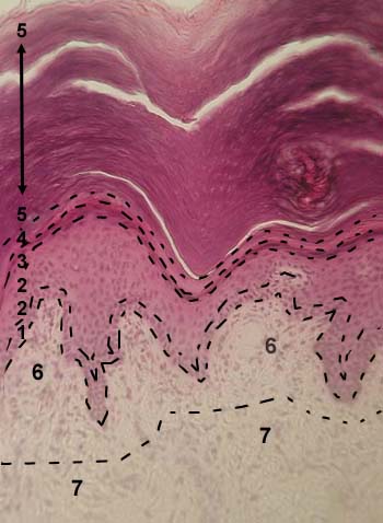
SKIN OF THE FINGER
Stained with haematoxylin and eosin

1 - basal layer of epidermis
2 - prickle cell layer (stratum spinosum) of epidermis
3 - granular layer of epidermis
4 - lucidar layer of epidermis
5 - cornified layer of epidermis
6 - papillary layer of dermis
7 - reticular layer of dremis

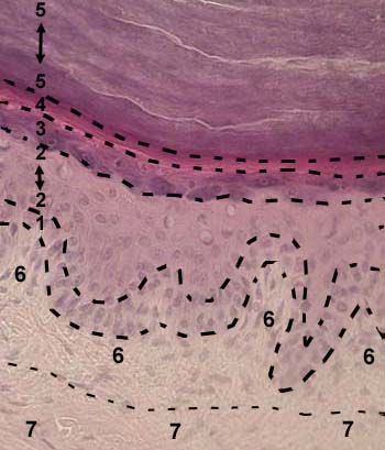
SKIN OF THE FINGER
Stained with haematoxylin and eosin

1 - basal layer of epidermis
2 - prickle cell layer (stratum spinosum) of epidermis
3 - granular layer of epidermis
4 - lucidar layer of epidermis
5 - cornified layer of epidermis
6 - papillary layer of dermis
7 - reticular layer of dremis


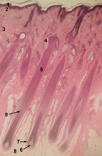
HAIR SKIN
Stained with haematoxylin and eosin

1 - epidermis
2 - papillary layer of dermis
3 - reticular layer of dremis
4 - sebaceous gland
5 - hair follicle
6 - dermal papilla
7 - hair bulb
8 - hypodermis
9 - hair root

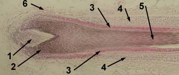
HAIR FOLLICLE
Stained with haematoxylin and eosin

1 - dermal papilla
2 - hair bulb
3 - inner root sheath
4 - outer root sheath
5 - hair root
6 - connective tissue sheath


__________________________________________________________________


URINARY SYSTEM

 
KIDNEY

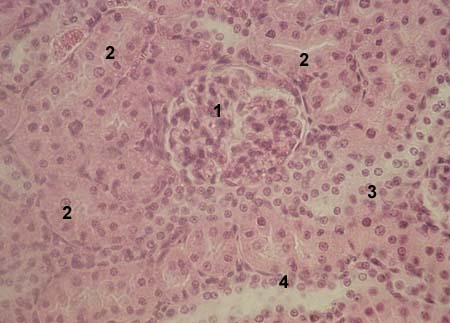
KIDNEY
     Stained with haematoxylin and eosin

1 - renal corpuscle
2 - proximal convoluted tubules
3 - distal convoluted tubules
4 - collecting tubule

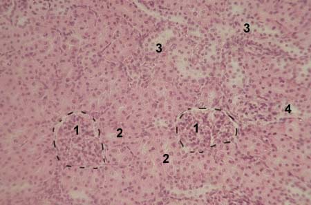
KIDNEY
cortex

     Stained with haematoxylin and eosin

1 - renal corpuscle
2 - proximal convoluted tubules
3 - distal convoluted tubules
4 - collecting tubule

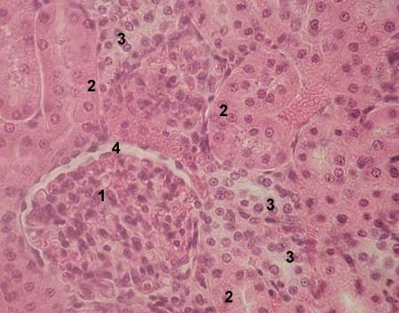
KIDNEY
cortex

     Stained with haematoxylin and eosin

1 - renal corpuscle
2 - proximal convoluted tubules
3 - distal convoluted tubules
4 - Bowman's capsulae space

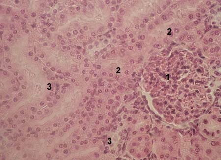
KIDNEY
cortex

     Stained with haematoxylin and eosin

1 - renal corpuscle
2 - proximal convoluted tubules
3 - distal convoluted tubules

KIDNEY
cortex

     Stained with haematoxylin and eosin

1 - renal corpuscle
2 - proximal convoluted tubules
3 - distal convoluted tubules
4 - collecting tubule

KIDNEY
medulla

     Stained with haematoxylin and eosin

1 - thick ascending limb of the loop of Henle
2 - thin descending limb of the loop of Henle
3 - collecting tubule

KIDNEY
medulla

     Stained with haematoxylin and eosin

1 - thick ascending limb of the loop of Henle
2 - thin descending limb of the loop of Henle
3 - collecting tubule


KIDNEY
medulla

     Stained with haematoxylin and eosin

1 - thick ascending limb of the loop of Henle
2 - interstitial connective tissue

KIDNEY
medulla

     Stained with haematoxylin and eosin

1 - thick ascending limb of the loop of Henle
2 - interstitial connective tissue

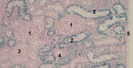
KIDNEY
absorbtion of dye by proximal convoluted tubules
     Stained with haematoxylin and eosin

1 - renal corpuscle
2 - proximal convoluted tubule
3 - distal convoluted tubule
4 - collecting tubule
5 - capsule of the kidney

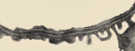
GLOMERULAR FILTER
(border between blood and primary urina)
    
Electron microscopy image

1 - capillary endothelium of the glomerulus
2 - podocyte pedicles, or secondary foot processes
     (inner layer of the glomerular (Bowman's) capsulae)
3 - glomerular basement membrane; there are 3 layers:
     middle layer is more dark (lamina densa), outher layers look
      lighter (laminae rara)


GLOMERULAR FILTER
    
Electron microscopy image


__________________________________________________________________


REPRODUCTIVE SYSTEM


MALE REPRODUCTIVE SYSTEM
TESTIS

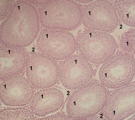
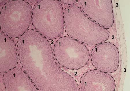
TESTIS
     Stained with haematoxylin and eosin

1 - seminiferous tubules
2 - interstitial connective tissue

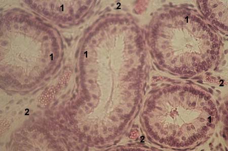
TESTIS
     Stained with haematoxylin and eosin

1 - seminiferous tubules
2 - interstitial connective tissue
3 - tunica albuginea

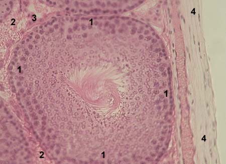
TESTIS
     Stained with haematoxylin and eosin

1 - seminiferous tubule
2 - interstitial connective tissue
3 - Leydig cells (interstitial cells)
4 - tunica albuginea

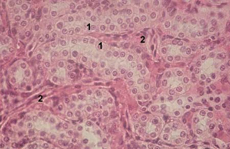
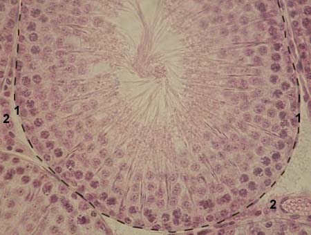
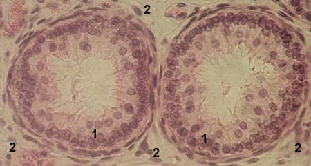
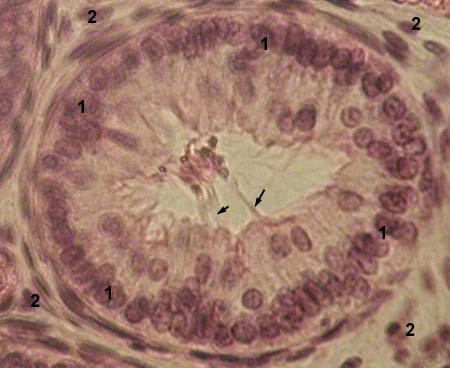
TESTIS
     Stained with haematoxylin and eosin

1 - seminiferous tubule
2 - Leydig cells


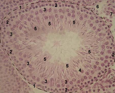
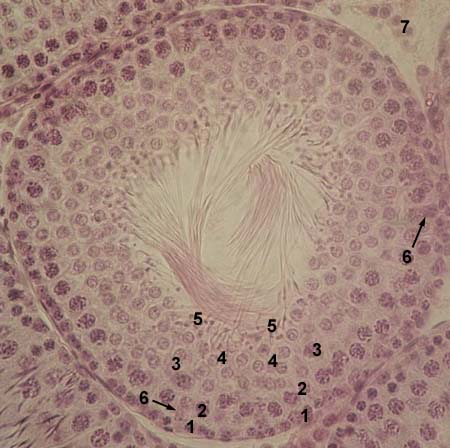
TESTIS
     Stained with haematoxylin and eosin

1 - seminiferous tubule
2 - spermatogonia
3 - primary spermatocytes
4 - secondary spermatocytes
5 - spermatids
6 - interstitial connective tissue
7 - Leydig cells

TESTIS
     Stained with haematoxylin and eosin

1 - seminiferous tubule
2 - spermatogonia
3 - primary spermatocytes
4 - secondary spermatocytes
5 - spermatids
6 - nuclei of Sertoli cells
7 - Leydig cells


__________________________________________________________________


MALE REPRODUCTIVE SYSTEM
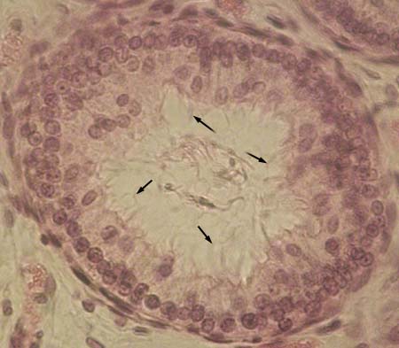
EPIDIDYMIS

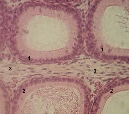
EPIDIDYMIS
ductulus of the head of the epididymis
(ductulus efferens)
     Stained with haematoxylin and eosin

1 - ductulus efferens
2 - interstitial connective tissue

EPIDIDYMIS
ductulus of the head of the epididymis
(ductulus efferens)
     Stained with haematoxylin and eosin

1 - ductulus efferens
2 - interstitial connective tissue

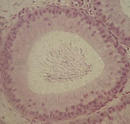
EPIDIDYMIS
ductulus of the head of the epididymis
(ductulus efferens)
     Stained with haematoxylin and eosin

1 - ductulus efferens
2 - interstitial connective tissue

  cilia of the epithelial cells
  are shown with arrows

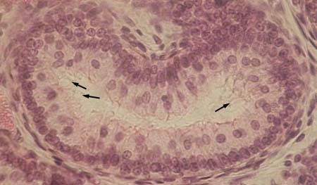
EPIDIDYMIS
ductulus of the head of the epididymis
(ductulus efferens)
     Stained with haematoxylin and eosin

  cilia of the epithelial cells
  are shown with arrows

EPIDIDYMIS
ductulus of the head of the epididymis
(ductulus efferens)
     Stained with haematoxylin and eosin

  cilia of the epithelial cells
  are shown with arrows

EPIDIDYMIS
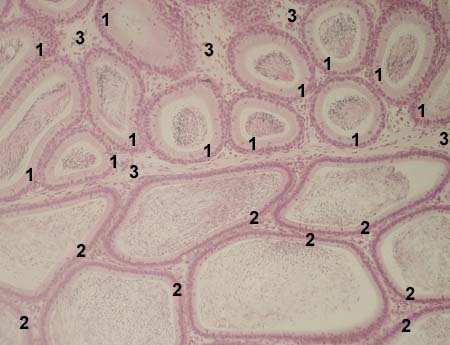
ductulus of the body and caudal pard of the epididymis
     Stained with haematoxylin and eosin

1 - proximal part of the ductulus of the body of the epididymis
2 - distal part of the ductulus of the body of the epididymis
3 - interstitial connective tissue


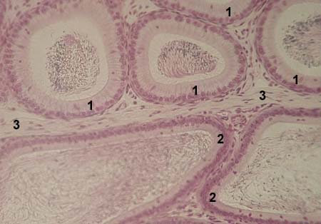
EPIDIDYMIS
ductulus of the body of the epididymis
     Stained with haematoxylin and eosin

1 - proximal part of the ductulus of the body of the epididymis
2 - distal part of the ductulus of the body of the epididymis
3 - interstitial connective tissue

EPIDIDYMIS
ductulus of the body of the epididymis
     Stained with haematoxylin and eosin

1 - proximal part of the ductulus of the body of the epididymis
2 - distal part of the ductulus of the body of the epididymis
3 - interstitial connective tissue

EPIDIDYMIS
ductulus of the body of the epididymis

     Stained with haematoxylin and eosin


__________________________________________________________________


MALE REPRODUCTIVE SYSTEM
PROSTATE GLAND

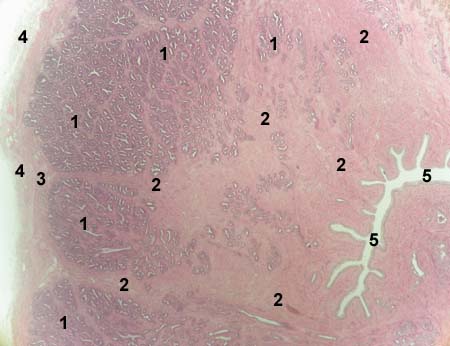
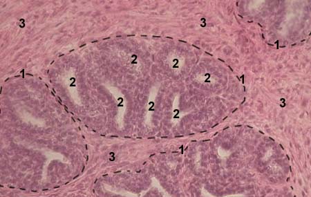
PROSTATE GLAND
     Stained with haematoxylin and eosin

1 - main prostatic glands
2 - stroma composed from smooth muscle cells and connective tissue
3 - stroma composed from smooth muscle cells and connective tissue
4 - fibro-elastic capsule
5 - prostatic part of the urethra

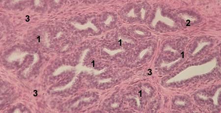
PROSTATE GLAND
     Stained with haematoxylin and eosin

1 - end secretory parts of main prostatic glands
2 - intralobular excretory duct
3 - stroma composed from smooth muscle cells and connective tissue

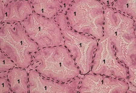
PROSTATE GLAND
     Stained with haematoxylin and eosin

1 - lobules of the main proststic glands
2 - end secretory parts of main prostatic glands
3 - stroma composed from smooth muscle cells and connective tissue

PROSTATE GLAND
     Stained with haematoxylin and eosin

1 - end secretory parts of main prostatic glands

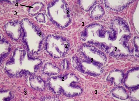
PROSTATE GLAND
     Stained with haematoxylin and eosin

2 - end secretory parts of main prostatic glands
3 - stroma composed from smooth muscle cells and connective tissue
4 - prostatic concretions in the end secretory parts of the glands


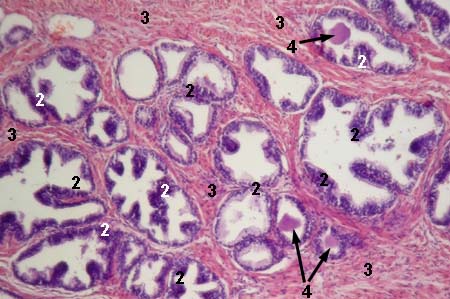
PROSTATE GLAND
     Stained with haematoxylin and eosin

2 - end secretory parts of main prostatic glands
3 - stroma composed from smooth muscle cells and connective tissue
4 - prostatic concretions in the end secretory parts of the glands

PROSTATE GLAND
transitional epithelium of the prostatic part of the urethra
     Stained with haematoxylin and eosin

1 - transitional epithelium
2 - tunica propria of the mucosa of 
      prostatic part of the urethra


__________________________________________________________________


FEMALE REPRODUCTIVE SYSTEM

 
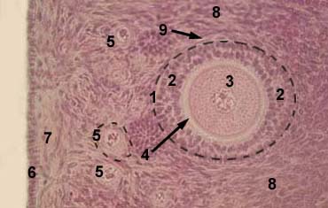
OVARY

OVARY
Stained with haematoxylin and eosin
general view

1 - cortex
2 - medulla
3 - area where primordial follicles are located
4 - primordial follicles

5 - secondary follicles
6 - tertiary follicles
7 - mature follicles (Graafian follicles)
8 - atretic follicles

cortex

cortex

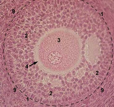
1 - secondary follicle
2 - follicular (granulosa) cells
3 - oocyte
4 - zona pellucida
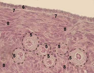
5 - primordial follicle

6 - simple cuboidal epithelium
      which covers the ovary
7 - tunica albuginea
8 - interstitial connective tissue
9 - theca

cortex
primary follicle

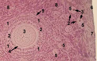
cortex
secondary follicle

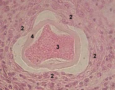
1 - primary follicle
2 - follicular (granulosa) cells
3 - oocyte
4 - zona pellucida
5 - primordial follicle
8 - interstitial connective tissue
9 - theca

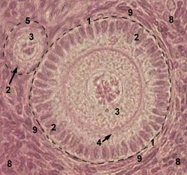
1 - secondary follicle
2 - follicular (granulosa) cells
3 - oocyte
4 - zona pellucida
5 - secondary follicles
8 - interstitial connective tissue
9 - theca


cortex
primordial follicles

cortex
atretic follicle

5 - primordial follicles
6 - simple cuboidal epithelium
7 - tunica albuginea
8 - interstitial connective tissue

2 - follicular cells with reduced
     volume of the cytoplasm
3 - wrinkled oocyte
4 - zona pellucida

cortex
corpus luteum

cortex
corpus luteum

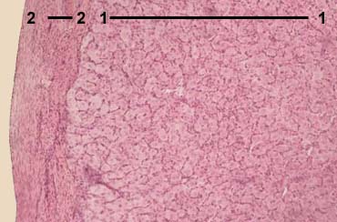
1 - parenchyma of the corpus luteum
2 - connective tissue capsule

5 - luteocytes
6 - interstitial connective tissue


__________________________________________________________________


FEMALE REPRODUCTIVE SYSTEM
UTERUS

UTERUS (proliferative phase)
    Stained with haematoxylin and eosin

1 - tunica mucosa (endometrium)
2 - tunica muscularis (myometrium)
3 - tunica serosa (perimetrium)
4 - functional layer of the endometrium
5 - basal layer of the endometrium
6 - blood vessels
7 - endometrial glands (in tunica propria
     of uterine mucosa)
8 - endometrial stroma (connective tuissue in
     tunica propria of uterine mucosa)
9 - epithelium of the endometrium

UTERUS (proliferative phase)
     Stained with haematoxylin and eosin

1 - tunica mucosa (endometrium)
2 - tunica muscularis (myometrium)
3 - tunica serosa (perimetrium)
4 - functional layer of the endometrium
5 - basal layer of the endometrium
6 - blood vessels
7 - endometrial glands (in tunica propria
     of uterine mucosa)
8 - endometrial stroma (connective tuissue in
     tunica propria of uterine mucosa)
9 - epithelium of the endometrium


__________________________________________________________________


FEMALE REPRODUCTIVE SYSTEM
MAMMARY GLAND

MAMMARY GLAND
(lactating)
     Stained with haematoxylin and eosin

1 - lobules of the gland
2 - end secretory units (alveoli, acini)
3 - intralobular conncective tissue
4 - interlobular excretory duct
5 - interlobular conncective tissue
6 - general excretory duct

MAMMARY GLAND
(lactating)
     Stained with haematoxylin and eosin

1 - lobule of the gland
2 - end secretory units (alveoli, acini)
3 - intralobular conncective tissue
4 - interlobular excretory duct
5 - interlobular conncective tissue
6 - interlobular excretory duct

MAMMARY GLAND
(lactating)
      Stained with haematoxylin and eosin

1 - lobules of the gland
2 - interlobular conncective tissue
3 - end secretory units (alveoli, acini)
4 - intralobular conncective tissue


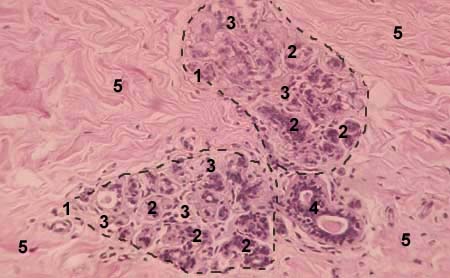
MAMMARY GLAND
(nonlactating)
     Stained with haematoxylin and eosin

1 - lobules of the gland
2 - precursors of end secretory units (alveoli)
3 - intralobular conncective tissue
4 - interlobular excretory duct
5 - interlobular conncective tissue

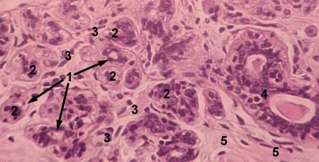
MAMMARY GLAND
(nonlactating)

     Stained with haematoxylin and eosin

1 - intralobular excretory duct
2 - precursors of end secretory units (alveoli)
3 - intralobular conncective tissue
4 - interlobular excretory duct
5 - interlobular conncective tissue


References: